Presentation
Cyclic lower abdominal pain. Left adnexal solid mass lesion on US exam.
Patient Data
Age: 35 years
Gender: Female
From the case:
Ovarian fibroma
![](https://prod-images-static.radiopaedia.org/images/63739699/74a17e1e8c1221689bf90d095602afde6b1d2cc4a0453ad6f7dd951239c586f9_thumb.jpeg)
![This study is a stack](/packs/stack-YQKLCKBI.gif)
![](https://prod-images-static.radiopaedia.org/images/63739719/0f92d30813f9a0f5a5a13024912a16f06313599151ea024f7511cffa94afb3f9_thumb.jpeg)
![This study is a stack](/packs/stack-YQKLCKBI.gif)
![](https://prod-images-static.radiopaedia.org/images/63739801/b98800fe9b80441a2d244775892ada1d639d9c28086403fe296d0182bc8ce5b2_thumb.jpeg)
![This study is a stack](/packs/stack-YQKLCKBI.gif)
![](https://prod-images-static.radiopaedia.org/images/63739745/195d60717628090372b7eabec479f7050d423e200acdb8fd6306a38ccfb927a4_thumb.jpeg)
![This study is a stack](/packs/stack-YQKLCKBI.gif)
![](https://prod-images-static.radiopaedia.org/images/63739824/8c25bb51336e586d7d21e9176b76c56ca5ef7ea315baa64d82e34b27a1142b1e_thumb.jpeg)
![This study is a stack](/packs/stack-YQKLCKBI.gif)
![](https://prod-images-static.radiopaedia.org/images/63739762/0d7627afffb73abefe29410916185decb5397bf118a9f06c2849334475a57d01_thumb.jpeg)
![This study is a stack](/packs/stack-YQKLCKBI.gif)
![](https://prod-images-static.radiopaedia.org/images/63739780/74624f97b585cbb0bdcebf1c3fe5f0bfda938554cf11691f8d83d3e1d3a809dc_thumb.jpeg)
![This study is a stack](/packs/stack-YQKLCKBI.gif)
![](https://prod-images-static.radiopaedia.org/images/63739848/2051bb67e13410be42d4d10297b2609fbde1f077491c1d09d78026f0495730e9_thumb.jpeg)
![This study is a stack](/packs/stack-YQKLCKBI.gif)
![](https://prod-images-static.radiopaedia.org/images/63739891/b99dd22f312b4e5c907ff2ab6c3e794d1ec27afb4299d08a2d3c509b1d6f81f3_thumb.jpeg)
![This study is a stack](/packs/stack-YQKLCKBI.gif)
![](https://prod-images-static.radiopaedia.org/images/63739910/7a0803def74b527a4356e2c9ff9de2f63c9c02a59057e49f99222157e5efe2f1_thumb.jpeg)
![This study is a stack](/packs/stack-YQKLCKBI.gif)
![](https://prod-images-static.radiopaedia.org/images/63740000/e90cf1ae1f3c388b9b4c71142636738c1a23a08e7a3fe421751d534def4fb9cc_thumb.jpeg)
![This study is a stack](/packs/stack-YQKLCKBI.gif)
Download
Info
![](https://prod-images-static.radiopaedia.org/images/63739699/74a17e1e8c1221689bf90d095602afde6b1d2cc4a0453ad6f7dd951239c586f9_big_gallery.jpeg)
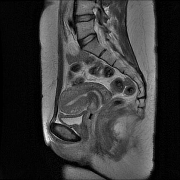
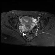
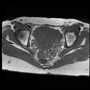
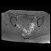
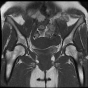
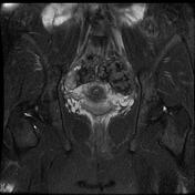
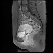
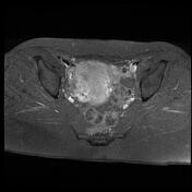
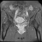
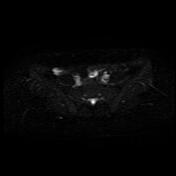
Well-defined left adnexal solid mass, measuring 20 x 30 x 40 mm. It appears of low signal intensity on T1, and very low signal intensity on T2 without obvious post-contrast enhancement. No evidence of water restriction is depicted on DWI images.
Case Discussion
MRI features are most consistent with the left ovarian fibroma. Ovarian fibroma is a benign tumour that arises from the ovary connective tissues. It is often difficult to differentiate from a pedunculated subserous fibroid.