
Presentation
Pelvic pain and palpable mass on physical exam.
Patient Data
Age: 55 years
Gender: Female
From the case:
Ovarian serous cystadenocarcinoma


Download
Info
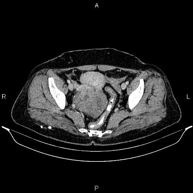
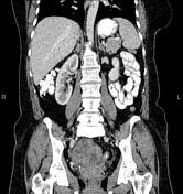
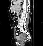
Two intramural masses 20 mm and 17 mm in diameters were seen in uterus inferring fibroids.
A large mass with lobulated margin and heterogeneous enhancement was seen behind uterus, measuring about 74×65×73 mm. The mass seems to originated from right ovary.
A little free fluid was seen in right adnexa.
Case Discussion
Pathology proven right ovarian serous cystadenocarcinoma which is the malignant form of ovarian serous tumor, the most common type of ovarian epithelial tumor. It is the most common type of ovarian malignancy.
No local invasion, no regional lymphadenopathy, and no metastasis.


 Unable to process the form. Check for errors and try again.