
Presentation
Work up for pelvic pain and fullness on physical exam.
Patient Data
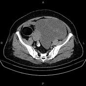

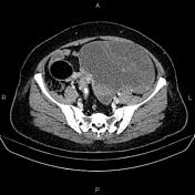
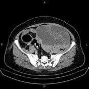

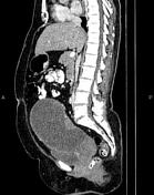

A 197×144×167 mm thick walled septated cystic lesion with enhancing solid components is seen at the left adnexa that displaces the urinary bladder and uterus to the right side.
A few enlarged lymph nodes are seen in bilateral parailiac regions with SAD less than 20mm.
Additionally, a 53×48 mm fat-containing mass with foci of marginal calcifications is seen at the right adnexa inferring ovarian dermoid cyst.
Inhomogeneously, the hepatic attenuation value is less than that of the spleen, suggesting uneven fatty liver.
The left kidney is relatively smaller than the right kidney and shows focal parenchymal thinning, particularly at the upper pole most consistent with scar. Additionally, a few small parapelvic cysts are seen in the kidneys.
Case Discussion
The patient underwent a total abdominal hysterectomy, and bilateral salpingo oophorectmy and histopathology evaluation confirmed left-sided ovarian serous cystadenocarcinoma also right-sided mature cystic ovarian teratoma without malignant transformation.


 Unable to process the form. Check for errors and try again.