
Presentation
Abdominal distension
Patient Data
Age: 20 years
Gender: Female
From the case:
Ovarian serous cystadenoma


Download
Info

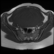
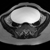
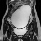
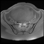
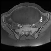
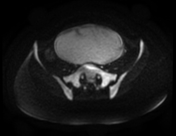
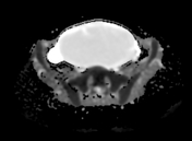
There is a large unilocular abdominopelvic cystic lesion, located in the midline arising from the left ovary with no solid component or internal septations. No restricted diffusion on DWI/ADC or abnormal enhancement on postcontrast sequences.
The ovary appears normal.
No pelvic ascites or enlarged lymph nodes.
Case Discussion
MRI features of a large unilocular cystic mass with no restricted diffusion and negative tumoral markers (in this case) in a young patient is suggestive of a benign ovarian tumor most likely a serous cystadenoma.


 Unable to process the form. Check for errors and try again.