
Presentation
Right lower anterior - lateral chest wall swelling - Few days. Referred for ultrasound to evaluate the swelling.
Patient Data
Age: about 20 - 25 yrs
Gender: Female
From the case:
Panniculitis - chest wall
Download
Info
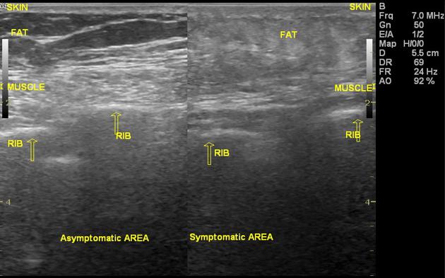
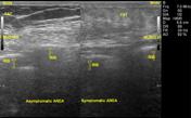
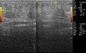
Subcutaneous fat shows increased echogenesity in the region of interest.
Approximate Size
Antero-posterior - 2.5 cm
Transverse - 10 cm
Craniocaudal - 4 cm
No collection was observed.
Doppler - No flow noted.
The underlying muscles are normal.
The underlying ribs show an intact anterior cortex.


 Unable to process the form. Check for errors and try again.