
Presentation
The patient presented with a chronic headache, bizarre behavior and aggression. No past medical history apart from controlled diabetes mellitus and hypertension.
Patient Data
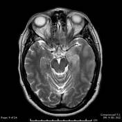


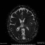
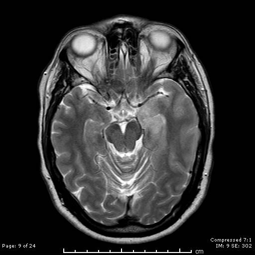
There is enlargement and increased T2 and FLAIR signal of the left medial temporal lobe. Increased T2 and FLAIR signal of the left insular cortex and temporoparietal cortex. Another lesion with similar abnormal signal is noted at the left thalamus. Also similar linear abnormal signal at the left side of the splenium of corpus callosum.
No significant peri-lesional edema, however, there are mild mass effects in the form of effacement of the related cortical sulci, mild compression of the third ventricle and slight encroachment on the left aspect of circum-mesencephalic cistern.
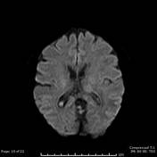
No evidence of diffusion restriction.
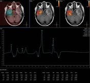
Intermediate TE MRS revealed reduction in NAA and marked elevation of Cho and Cr peaks.
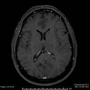
The above lesions did not show significant enhancement after contrast administration.
Case Discussion
Paraneoplastic limbic encephalitis is associated with various malignant neoplastic lesions, most commonly bronchial carcinoma and non-Hodgkin lymphoma.
In this case, the patient had gastric non-Hodgkin lymphoma with no alarming gastrointestinal manifestations.
The patient started to have aggressive bizarre attitude and persistent headache. MRI was delayed for three months after the beginning of the symptoms as the patient was claustrophobic. After MRI, the patient has not sought medical advice and she died a month later.
An autopsy has proven gastric non-Hodgkin lymphoma.


 Unable to process the form. Check for errors and try again.