
Presentation
Young male with a three month history of a right parotid lump. No facial weakness. No palpable cervical nodes.
Patient Data


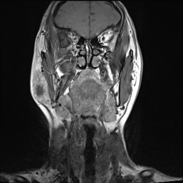
A well defined low T1, T2 hyperintense homogeneously enhancing mass in the superficial lobe of the right parotid gland is present. The medial aspect of the mass abuts the retromandibular vein, with no extension into the deep lobe or extra-capsular extension. No intra-parotid lymphadenopathy.
Borderline right submandibular space lymph node.
Case Discussion
The patient went on to have a FNAC.
Cytology
Multiple smears prepared from the FNA of the right parotid gland show ductal epithelial and myoepithelial cells situated in a fibromyxoid stroma. Some of the cells show plasmacytoid pattern. The appearances are those of a pleomorphic adenoma.
Discussion
Pleomorphic adenomas account for 70-80% of benign salivary gland tumors and are especially common in the parotid gland, and are most often located in the superficial lobe.
Three histological types have been described:
- myxoid (hypocellular) - which is the most frequently observed.
- cellular
- classic
The myxoid type has a very hyperintense appearances as in this case.


 Unable to process the form. Check for errors and try again.