
Presentation
Left parotid pain
Patient Data
Age: 50 years
Gender: Male
From the case:
Parotid sialolithiasis


Download
Info

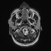
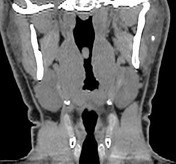
No definite lesion could be seen on the left parotid gland.
From the case:
Parotid sialolithiasis

Download
Info
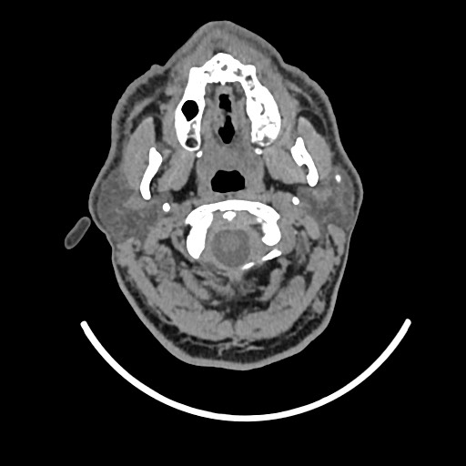
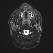
A small intraglandular stone is noted at the anterior part of the left parotid gland with no associated duct dilatation.
Case Discussion
Small sialolithiasis may not be identified on MRI especially if there is no ductal dilatation. CT is the modality of choice for sialolithiasis.


 Unable to process the form. Check for errors and try again.