
Presentation
Persistent pain and periodic swelling after trauma.
Patient Data
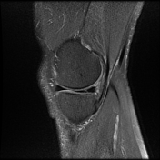


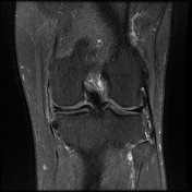
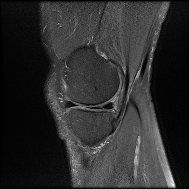
An oblique radial medial meniscal tear of the free edge is visible at the junction between the body and posterior horn. The tear results in a marching cleft sign on sagittal images and has a curvilinear course on axial images, in keeping with a parrot beak tear.
Case Discussion
Care must be taken to describe the configuration of a meniscal tear, especially if surgery is considered. The so-called parrot beak tear is a radial-oblique tear of the meniscal free edge. Its curvilinear course may be nicely appreciated on axial images if the cross-section runs through the meniscus. On sagittal images it produces a marching cleft sign; however, this appearance is not specific as it can also be present is simple radial tears located at the junction of the meniscal body and horn. Therefore, calling a tear a parrot beak is not recommended on the base of this finding only.


 Unable to process the form. Check for errors and try again.