
Presentation
Pregnant female 16 weeks, antenatal ultrasound screening
Patient Data
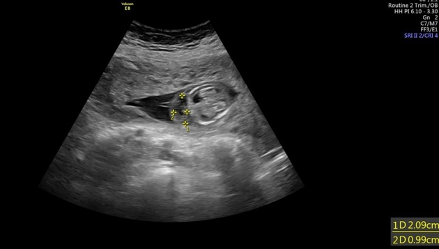
Large anterior placenta with multiple variable-sized cysts.
Single viable intra-uterine pregnancy with evidence of marked fetal ascites, subcutaneous edema, and thickened nuchal translucency.


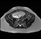
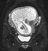
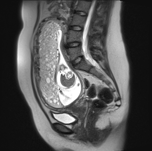
The placenta is enlarged (relative to the size of the uterine cavity) and anteriorly located, reaching the fundus superiorly and the edge of internal cervical os inferiorly. There are extensive internal cystic spaces characteristic of 'molar placenta'.
The amniotic cavity shows an internal fetus with evidence of hydrocephalus and marked ascites.
The outer myometrium appears smooth with no evidence of gross invasion. However, it appears hypervascular with extensive tortuous vessels.
Case Discussion
Here we show a case of partial hydatidiform mole, associated with molar placenta and alive fetus with hydrocephalus and marked ascites.
The total beta HCG level was very high (175,969 mIU/mL). MRI has an important role in the exclusion of myometrial invasion that was negative in this case. However serial beta HCG level follow up is recommended after evacuation to exclude the possibility of persistent trophoblastic disease and invasive mole.
Ultrasound contribution by Dr Samar Fathy.


 Unable to process the form. Check for errors and try again.