
Presentation
Pain and swelling of the knee, after a lateral patellar dislocation.
Patient Data

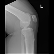
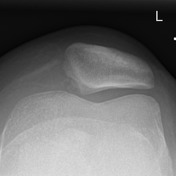

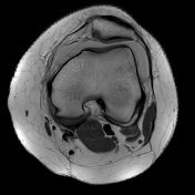
Findings:
a small avulsed fragment at the medial rim of the patella
Impression:
- medial avulsion fracture of the patella


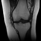
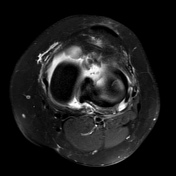
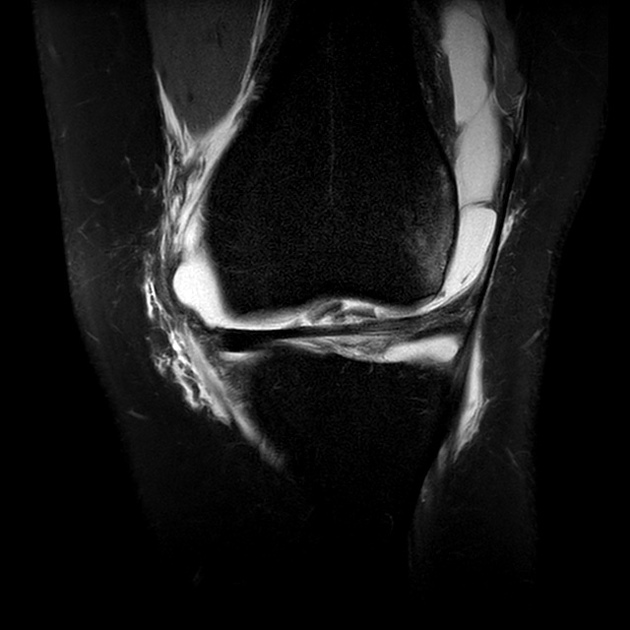
Findings:
large knee joint effusion
Intercondylar region:
- unremarkable anterior and posterior cruciate ligaments
- two loose chondral fragments between the anterior transverse meniscal ligament and the intercondylar groove
Medial compartment:
- unremarkable cartilage and medial meniscus
- soft tissue edema
- thickening of the superficial part of the medial collateral ligament small tear within the proximal part
Lateral compartment:
- bone contusion of the anterolateral rim of the lateral femoral condyle
- extensive chondral lesion of the antero- and centrolateral femoral cartilaginous zone with delamination
- normal lateral collateral ligament and posterolateral corner
Patellofemoral compartment:
- medial patellofemoral ligament tear at the patellar and femoral attachment with nondisplaced osteochondral avulsion fracture at the inferomedial patella margin
- fluid extending into the vastus medialis myotendinous junction
- shallow trochlear groove
- Hoffa fat pad edema
Impression:
Typical findings of prior lateral patellar dislocation:
- medial patellofemoral ligament tear at the patellar and femoral attachment with osteochondral avulsion fracture at the inferomedial patellar margin
- two intraarticular chondral fragments
- extensive chondral lesion of the antero- and centrolateral femoral condyle
- traumatic changes of the Hoffa fat pad
- decreased trochlear depth indicating trochlear dysplasia

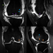
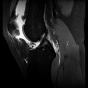
Key findings:
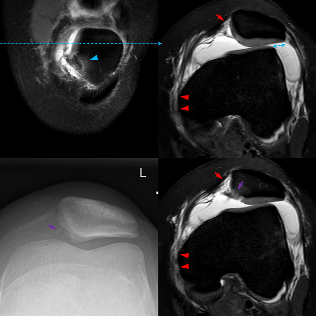
bone bruises of inferomedial patella (blue arrowhead) and the anterolateral femoral condyle (blue arrows)
patellar lateralization (blue double arrow)
medial patellar avulsion fracture with an unstable osteochondral fragment (purple arrow)
medial patellofemoral ligament with a partial tear of the patellar insertion (red arrow) and disruption of the femoral attachment (red arrowheads)
edema and myotendinous tear of the vastus medialis oblique (VMO) (green arrow)
two chondral fragments (purple arrowheads) between the anterior transverse meniscal ligament and the intercondylar groove
chondral lesion of the antero- and centrolateral femoral condyle (orange arrowheads) with delamination
Assessment for risk factors:
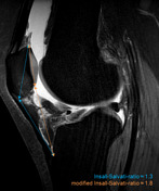
Insall-Salvati index ≈ 1,3 - borderline
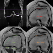
decreased trochlear depth (<3mm)
lateral trochlear inclination (LTI) and trochlear facet ratio within normal limits
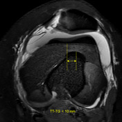
TT-TG < 15mm
Case Discussion
This case illustrates findings typical for prior lateral patellar dislocation 1:
- bone bruises at the typical locations at the inferomedial patella and the lateral femoral condyle
- injury of the medial stabilizers including the medial patellofemoral ligament, the vastus medialis oblique (VMO) and medial patellar retinaculum
- patellar lateralization
It also shows findings that can be associated with this condition 1-5:
- osteochondral and chondral injuries of the at the inferomedial patella and the lateral femoral condyle with chondral delamination
- intra-articular loose chondral fragments
- posttraumatic injury of the Hoffa fat pad
There is also a decreased trochlear depth indicating trochlear dysplasia, which is a risk factor for patellofemoral instability 1,2.
The patient underwent arthrotomy with a removal of the loose intra-articular chondral fragments, lavage and debridement of the patellar and lateral femoral condyle chondral lesions and refixation of the medial patellofemoral ligament.


 Unable to process the form. Check for errors and try again.