
Presentation
A large palpable mass proximal to the knee joint. Acute pain due to recent trauma.
Patient Data
Age: 20 years
Gender: Female
From the case:
Pedunculated osteochondroma with fracture
Show annotations
Download
Info
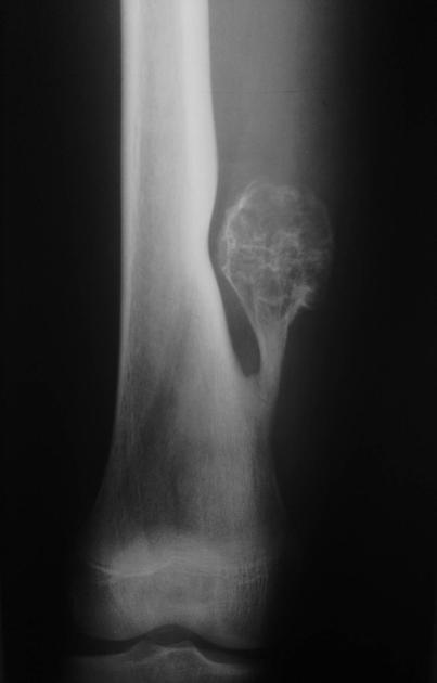
A pedunculated bony lesion is seen in the right knee's metaphyseal region projecting away from the epiphysis. There is a mild broadening of the metaphysis from which it arises. The cartilage cap is also noted with rings and arcs calcifications.
An undisplaced fracture through the neck is noted.
Case Discussion
The findings of the radiograph are consistent with pedunculated osteochondroma. The femur (especially the distal part) is the most common site of osteochondroma.
Case courtesy Prof. Saeed Rad, Tabriz, Iran.


 Unable to process the form. Check for errors and try again.