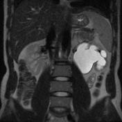
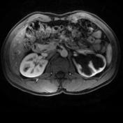
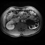
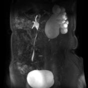
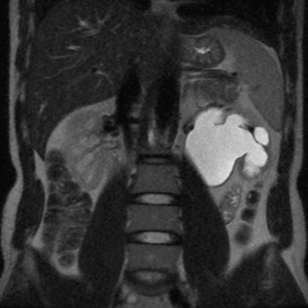
Q: What is the cause of left sided hydronephrosis?
show answer
A: Pelviureteric junction (PUJ) obstruction.
Q: In the above study, what additional protocol would have helped in knowing whether it is complete or partial obstruction?
show answer
A: Administration of furosemide (0.5-1mg/kg) during the study, may provoke excretion of contrast through left ureter in partial obstruction. However, complete obstruction would not have any passage of contrast even after provocation.
Q: In the above case, if calyces were dilated with normal pelvis, what would have been possible diagnosis?
show answer
A: Congenital megacalycosis.
Q: What would be possible causes of a ballooned renal pelvis with essentially normal calyceal system?
show answer
A: Extrarenal pelvis is a common variant.
Q: When found incidentally in an adult what are some of the underlying aetiologies which should be considered?
show answer
A: Congenital PUJ obstruction often remains asymptomatic, and as such is a likely diagnosis even in adults. Aberrant vessels, kinks or a band (e.g. retroperitoneal fibrosis) may cause extrinsic PUJ anomaly. Tumours or even infection (e.g. TB) at the PUJ can also cause localized obstruction.
Q: If while performing a plain film IVP a linear filling defect is seen at the pelviureteric junction what is the most likely cause? What simple technique can confirm this?
show answer
A: Extrinsic vascular compression is common, with or without renal pelvis prominence. Sometimes a prone film helps as the extrinsic compression is relieved.


 Unable to process the form. Check for errors and try again.