
Presentation
Sudden onset left iliac fossa pain.
Patient Data


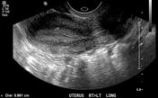

Two uterine cavities present, separated by a septum which continues into the cervix. The 3D coronal reformat unfortunately excludes the serosal fundal contour, however there is contiguous myometrium extending at least to the level of the cornua. Therefore if a fundal dip is present, it is likely to be shallow and hence uterine morphology is most likely septate, or borderline bicornuate.
Secretory endometrium, measuring 9mm on the right and 11mm on the left.
Normal ovaries with a corpus luteum on the left.
There is marked dilatation of the right renal pelvis, without evidence of hydroureter. There were no symptoms pertaining to the right kidney and this is most likely longstanding. Further evaluation with CT was undertaken to assess the renal tract.

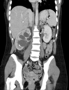
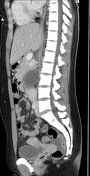

Gross dilatation of the right pelvicalyceal system with the transition point at the level of the pelviureteric junction. Right ureter is not dilated. There is no evidence of an obstructing calculus at the PUJ, though two intrarenal non obstructing stones are noted within the right lower pole calyces.
There is right renal cortical thinning and a global decrease in enhancement of the right renal parenchyma compared to the left.
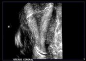
Two uterine horns are noted with only a small (approximately 5mm) dip of the serosal fundal contour - confirming septate uterine morphology.
Incidental right gluteus minimus lipoma.
Case Discussion
Two findings with CT and ultrasound correlation: a right PUJ obstruction and a septate uterus.
Mullerian duct anomalies are frequently associated with anomalies of the renal tract.


 Unable to process the form. Check for errors and try again.