Patient Data
Note: This case has been tagged as "legacy" as it no longer meets image preparation and/or other case publication guidelines.

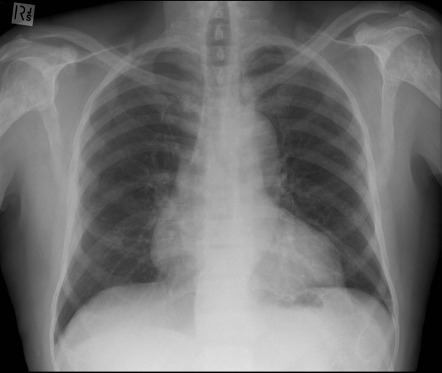
Plain film of the chest reveals multiple sclerotic bone metastases involving the bilateral scapula, humerus and ribs, in keeping with a diagnosis of metastatic prostatic cancer.
Incidental note is made of a prominent right heart border.

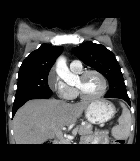
Incidental note is made of a prominent right heart border which, on review of the CT imaging, is shown a well-defined non-enhancing fluid-attenuated cyst, separated from the normal myocardium and heart chambers.
Case Discussion
Plain film of the chest reveals multiple sclerotic metastases involving the bilateral shoulders and ribs, in keeping with a diagnosis of metastatic prostatic cancer.
Incidental note is made of a prominent right heart border which, on review of the CT imaging, is shown to be secondary to a pericardial cyst.


 Unable to process the form. Check for errors and try again.