
Presentation
Nausea, vomiting, and epigastric pain. Refers to medical care for about 10 days for the same symptoms.
Patient Data


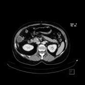
CT image shows faint signals of acute cholecystitis with calcified micro gallstones, wall thickening and pericholecystic fat stranding. A small collection is identified in lower position, near lateroconal fascia, with capsular enhancement.

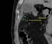
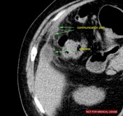

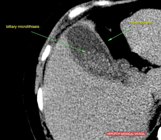
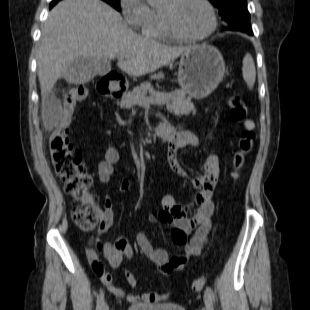
In these images is better seen the biliary microlithiasis into gallbladder as well it`s wall thickened (3.7 - 4.0 mm - better demonstrated on ultrasound study). It is identified the communication path between gallbladder and fluid collection.
Case Discussion
About 3 to 19% of acute cholecystitis are complicated with an abscess formation. Normally it's presented as small fluid collections near/adjacent to gallbladder with rim-enhancement, it can be also extended into the adjacent hepatic parenchyma.


 Unable to process the form. Check for errors and try again.