
Presentation
Left sciatica.
Patient Data


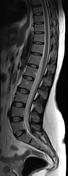
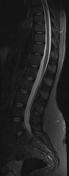
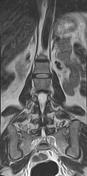
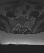
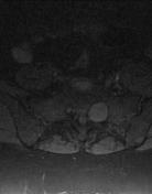
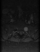
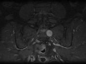
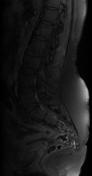
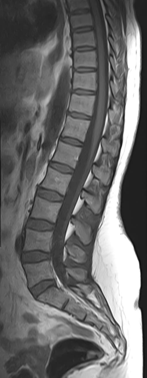
The MRI sequences demonstrate a well-defined ovoid mass 25 x 23 x 20 mm, centered on the left first sacral foramen which is enlarged. It elicits a low signal on T1, low signal centrally with hyperintense rim peripherally on T2 "target sign". The postcontrast sequences show homogeneous enhancement.
Case Discussion
MRI features of a well-defined mass enlarging the first left sacral foramen with "target sign" on T2 and homogeneous enhancement, suggestive of a peripheral nerve sheath tumor of S1 nerve root.
The "target sign" is highly suggestive of neurofibroma. It is thought to be due to the presence of collagenous stroma in the central area. Occasionally also can be seen in schwannomas and malignant peripheral nerve sheath tumors.


 Unable to process the form. Check for errors and try again.