
Patient Data
The patient had undergone a hip hemi-arthroplasty for a subcapital fracture of the neck of femur which had external rotation deformity in 2020.

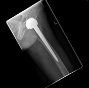
An AP pelvis projection was performed to include the entire implant. A turned lateral was also performed. Status post open reduction and internal fixation with application of a bipolar hemiarthroplasty of the left hip joint demonstrated satisfactory alignment. No evidence of a new fracture was noted.
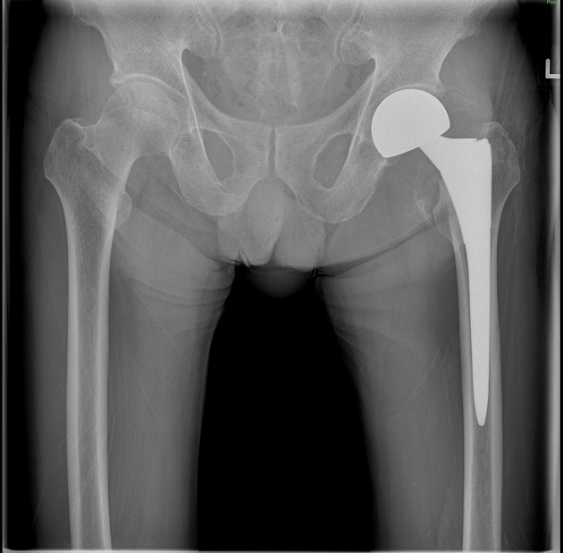
Patient presented at the A&E post fall, with pain over the left thigh in 2022.


An AP pelvis radiograph was attained, along with a horizontal beam lateral due to patient's history of trauma.
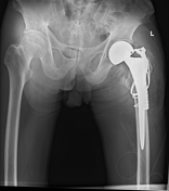
Reference made to prior radiograph. Status post left total hip replacement. There is a mildly displaced periprosthetic fracture around the subtrochanteric region of the left femur.
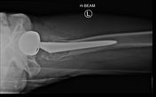
The patient was sent for an emergency operation.

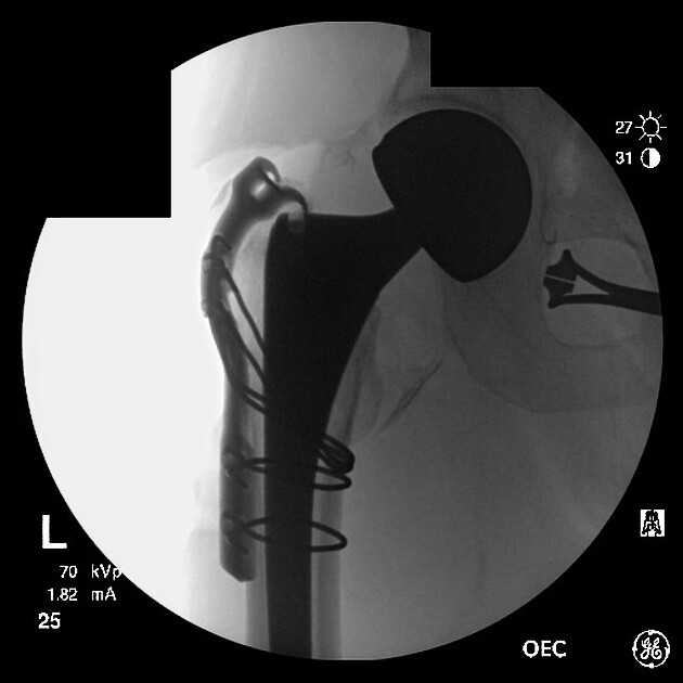
Integral long greater trochanteric reattachment device with 4 cables was inserted, position was satisfactory under fluoroscopy. The image orientation was of the surgeon's preference.
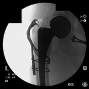
The day after, post operative radiographs were performed for the patient.
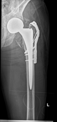
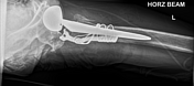

An AP pelvis was performed, with an additional AP hip for a better demonstration of the new implant. A lateral horizontal beam hip was also performed for the comfort of the patient. The left hip prosthesis with cerclage wires noted and early postoperative changes around the left hip and upper thigh were noted.


 Unable to process the form. Check for errors and try again.