
Presentation
Abdominal pain and weight loss.
Patient Data

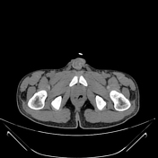

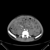
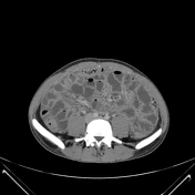
Diffuse smooth enhancing thickening of the peritoneal reflections and micro-nodular greater omentum and mesentery, abdominopelvic fluid density non-loculated ascites of a moderate amount.
No terminal ileum or caecal wall thickening, hyperaemia, or stricture. No feature of intestinal obstruction, bowel perforation, fistulae, abscesses, and/or haemorrhage
Few inguinal and mesenteric lymph nodes all are moderately enhancing, non-enlarged, with no necrosis, matting, or calcification.
Case Discussion
The patient had undergone appendicectomy one month ago, and the caecal biopsy showed chronic inflammatory infiltrate with multinucleated giant cells, however, the IGRA test was negative and no further workup was done.
CT findings of peritonitis likely tuberculous (the tumour marker results were negative), however the histopathological/laboratory assessment of the fluid aspirate, omental biopsy (preferably ultrasound guided), and culture are required to confirm the presence of caseating granulomas and AFB before commencing treatment.


 Unable to process the form. Check for errors and try again.