
Presentation
TIA
Patient Data
Note: This case has been tagged as "legacy" as it no longer meets image preparation and/or other case publication guidelines.
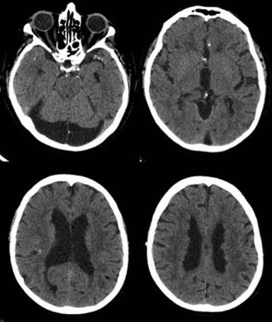
Non-contrast CT demonstrates nodularity of the ependymal surface of the ventricles giving it a lumpy bumpy appearance. Background of intracranial arterial calcifications and periventricular hypodensities which may represent microangiopathic changes. Posterior fossa cystic lesion of CSF attenuation and mild regional mass effect is probably either an arachnoid cyst or variant mega cisterna magna.
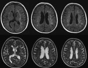

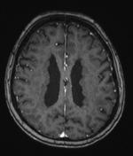
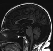

Extensive subependymal nodules line the walls of the lateral ventricles. These nodules follow gray matter signal intensity on all sequences and appearance is typical of periventricular nodular heterotopia.
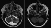
Background of semi-confluent periventricular and external capsule white matter hyperintensities consistent with microangiopathy. Incidental finding of a posterior fossa arachnoid cyst with marginal regional mass effect. Partial empty sella.
Case Discussion
Periventricular nodular heterotopia is a common cortical malformation in adults. Subependymal nodules of gray matter line the lateral walls of the ventricles and follow gray matter density on CT and signal intensity on MRI. These nodules do not enhance on post-contrast examination. When periventricular nodular heterotopia is recognized in female patients of reproductive age, genetic counseling is advised as FLNA x-linked dominant mutation is fatal to affected male resulting in perinatal/in utero demise.


 Unable to process the form. Check for errors and try again.