
Presentation
Intermittent high dysphagia for solids, and regurgitation.
Patient Data
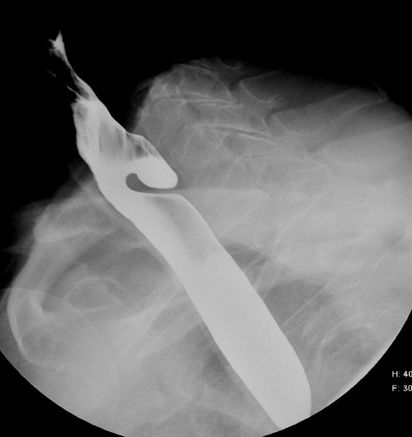
Left-sided 3 cm pharyngeal pouch arising at the C7 level, retaining contrast throughout the examination.
Normal oesophageal and gastric contrast transit (not included).
The patient was later listed for surgical repair of the pouch. Perforation was noted intraoperatively, and the next day the patient was febrile and tachycardic. CT performed to assess for collection/mediastinitis.
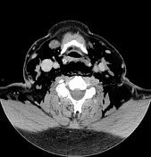

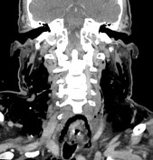
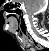
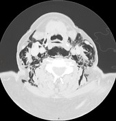
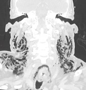
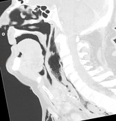
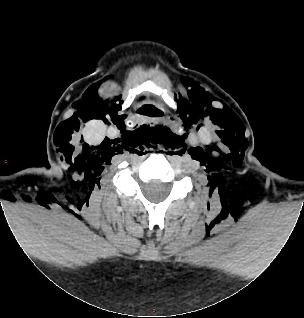
Extensive surgical emphysema in the soft tissues of the neck, with a large pocket in the retropharyngeal space. The gas surrounds the oesophagus, tracking into the mediastinum.
5 x 3 cm air and fluid filled collection posterior to the oesophagus compatible with a clipped pharyngeal pouch.
Gas and inflammatory stranding in the mediastinum, but no organised collection.
Case Discussion
The majority of pharyngeal pouches are managed conservatively, with surgery considered for symptomatic patients. Endoscopic repair is a common route, although there is a reported 2-6% rate of intraoperative perforation.


 Unable to process the form. Check for errors and try again.