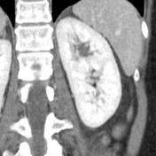
Pheochromocytoma and duplex system

Case contributed by Frank Gaillard
Diagnosis almost certain

Patient Data

Age: Adult
This study is a stack
Axial
non-contrast
This study is a stack
Coronal
non-contrast
This study is a stack
Axial renal
parenchymal phase
This study is a stack
Coronal renal
parenchymal phase
This study is a stack
Axial renal
excretory phase
This study is a stack
Coronal renal
excretory phase
This study is a stack
Coronal renal
parenchymal phase
Faceless
kidney
Download
Info

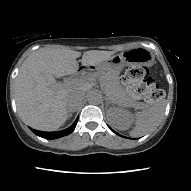
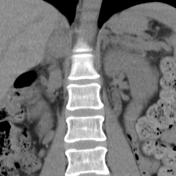
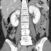
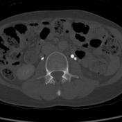
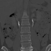
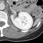
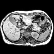
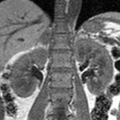
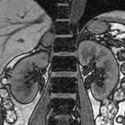
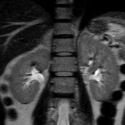
Duplex collecting system on the left, down to mid ureter, with axial image between systems demonstrating a faceless kidney sign. On the right, a heterogeneous adrenal mass is present. 

This study is a stack
Axial T1
in-phase
This study is a stack
Axial T1
out-of-phase
This study is a stack
Coronal T1
in-phase
This study is a stack
Coronal T1
out-of-phase
This study is a stack
Axial
T2
This study is a stack
Coronal
T2
Download
Info

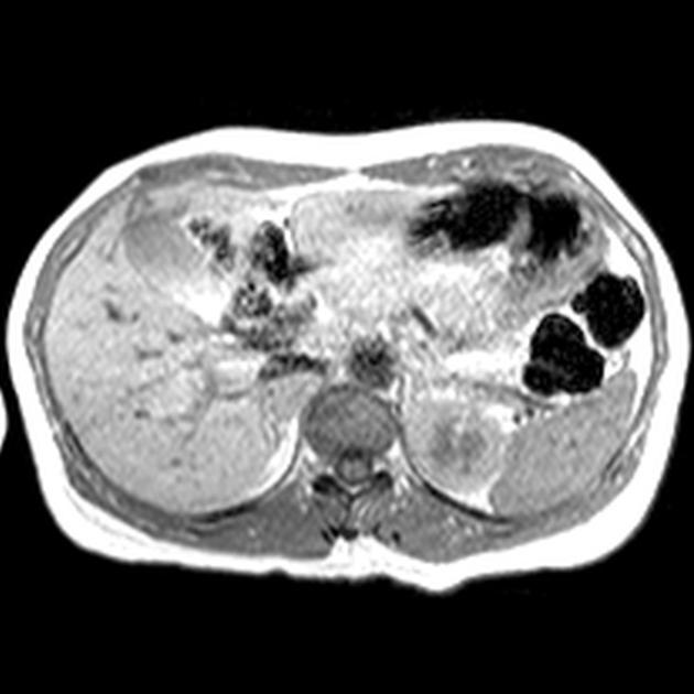
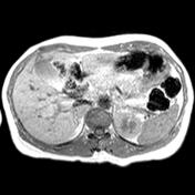
The right adrenal mass does not show signal-loss on out of phase imaging. 

Case Discussion

The right-sided adrenal mass went on to be shown histologically to be a pheochromocytoma

How to use cases

You can use Radiopaedia cases in a variety of ways to help you learn and teach.

Creating your own cases is easy.

:

Updating… Please wait.

 Unable to process the form. Check for errors and try again.

 Thank you for updating your details.