
Presentation
Knee joint pain, swelling and limitation of movement over the last 3 months
Patient Data
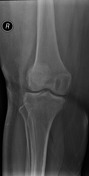
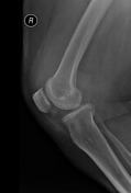
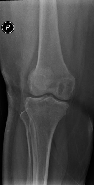
An osteolytic lesion is noted at the medial femoral condyle associated with large amount of knee joint effusion. The joint space is preserved.


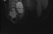

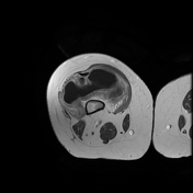
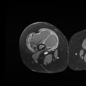
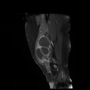
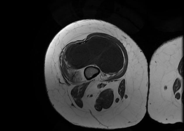
Diffuse synovial thickening that forms intra articular masses, the largest one is irregular in shape, is noted anterior to the medial femoral condyle and displaying low signal on T2 and T2* sequences. Large amount of knee joint effusion. Synovial enhancement following contrast administration is seen.
Case Discussion
The characteristic low T2 signal intensity of the nodular synovial thickening and masses is suggestive of pigmented villonodular synovitis (PVNS) due to hemosiderin deposition from repeated hemorrhage. A biopsy was taken from the sizable mass anterior to the medial femoral condyle and confirmed the diagnosis.
Editor's note: Per the 2020 WHO Soft Tissue and Bone Tumors Classification (5th ed.), the recommended terminology is tenosynovial giant cell tumor with pigmented villonodular synovitis no longer recommended (although remains in common use).


 Unable to process the form. Check for errors and try again.