
Presentation
Asymptomatic red nodule located on the head.
Patient Data
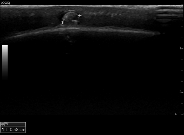
Sonography reveals a solid, oval-shaped subdermal mass with well-demarcated borders and a diameter of approximately 10 mm, above the galea capitis. The lesion is hypoechoic containing matrix of round calcification, poorly vascularized with peripheral hypoechoic rim.
Histology
Macroscopically, the mass measure approximately 10 x 5 mm without invasion into adjacent structures. Microscopic examination reveal a central area of calcified material measure up to 3 mm in diameter with epithelial cells surrounded by stromal cells and blood vessels. The specimen is consistent with pilomatricoma.
Case Discussion
Pilomatricoma is typically an isolated benign tumor of the hair follicle matrix. Pilomatricomas are generally found in the head and neck and, upper extremities, rarely on the chest, or lower extremities.


 Unable to process the form. Check for errors and try again.