
Presentation
Incidental finding.
Patient Data
Age: Adult


Download
Info
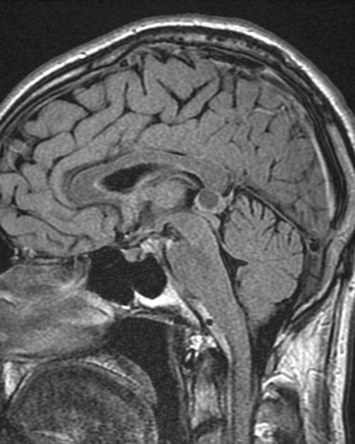
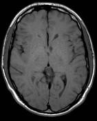
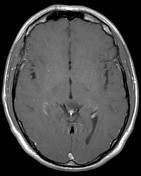
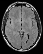
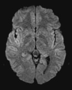
MRI of the brain demonstrates a small (<10mm) cystic lesion in the pineal region. It has high T2 and low T1 signal. On FLAIR it mostly attenuates, and there is no restricted diffusion on DWI. Following administration of contrast there is no solid enhancement.
Case Discussion
This case demonstrates typical appearances of an Incidental pineal cyst, which are found in up to 5% of all MRI brains, and as such pose a problem to deciding which should be followed up.
In general large size (>10-12mm) or any atypical features warrant followup to ensure that they do not represent a cystic pineal neoplasm (e.g. pineocytoma).


 Unable to process the form. Check for errors and try again.