
Presentation
2 months of severe occipital headache.
Patient Data


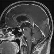


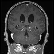
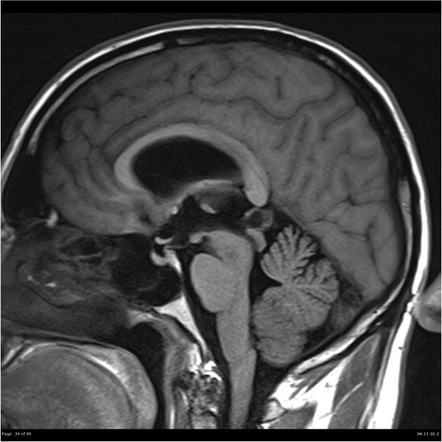
A lobulated 14 x 8 mm pineal cyst projects anteriorly to obstruct the upper margin of the cerebral aqueduct, with mild to moderate hydrocephalus and mild periventricular (transependymal) signal abnormality.
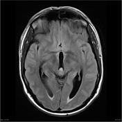
Several small T2 hyperintense lesions, measuring up to 5 mm diameter inferiorly in the right pre-central gyrus, are in keeping with chronic small vessel ischemic change, a little greater than expected for the patient's age, with no associated diffusion restriction or magnetic susceptibility. The remainder of the study is unremarkable.

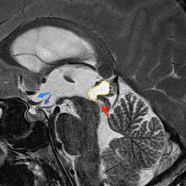
A cystic pineal region mass (yellow dotted line) has prolapsed anteriorly and obstructed the superior aspect of the aqueduct (red). Hydrocephalus is present with inferior bowing of the floor of the third ventricle and ballooning of the supraoptic recess (blue) and infundibular recess (purple).
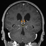
As is the cases with all pineal gland masses the internal cerebral veins (orange) are located above the mass.
The patient went on to have endoscopic resection of the mass.
Histology
MICROSCOPIC DESCRIPTION: Serial sections show a small fragment of glial tissue which incorporates a slit-like space. This is lined by a more compact layer of glial tissue staining strongly for GFAP but with focal staining for epithelial membrane antigen. Scattered single siderophages are noted in tissue adjacent to the space and there are also scattered Rosenthal fibers. No evidence of tumor is seen. No pineal parenchymal tissue is identified. The features are of pineal cyst/pineal glial cyst.
FINAL DIAGNOSIS: Pineal cyst
Case Discussion
Although pineal cysts are very common, and usually asymptomatic they can result in obstructive hydrocephalus, usually due to compression of the tectum. In this case the cyst has prolapsed into the aqueduct.


 Unable to process the form. Check for errors and try again.