
Patient Data
Age: 30 years
Gender: Male
Download
Info
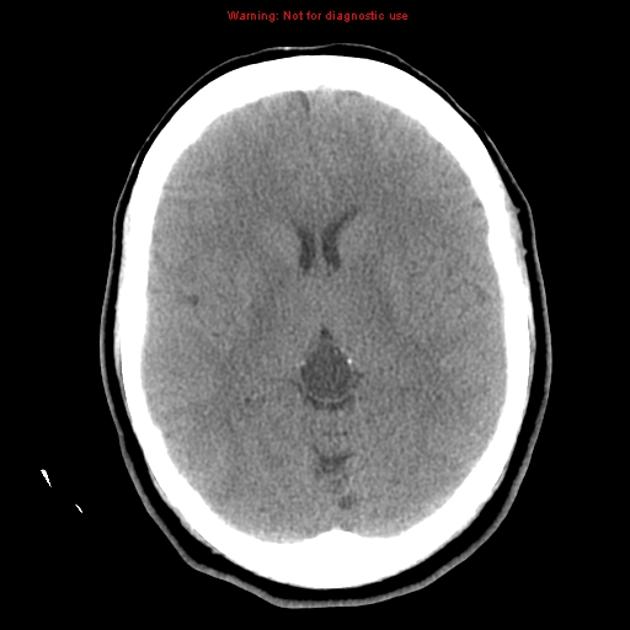
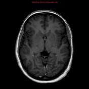
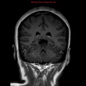
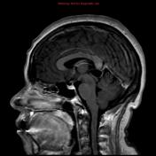
A well-defined cystic lesion in the pineal body containing scattered calcified foci.


Download
Info
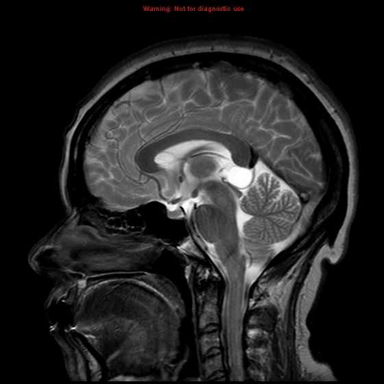
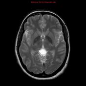
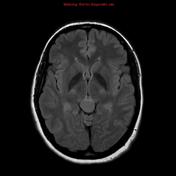
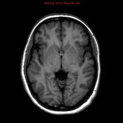
The lesion is of low-signal intensity on T1 and high signal intensity on T2 as well as intermediate signal intensity on FLAIR. No restricted diffusion is demonstrated. Mild linear enhancement is demonstrated after gadolinium injection. No nodular enhancement or soft tissue elements.
Case Discussion
This case illustrates the appearances of a large pineal cyst with fluid which does not fully attenuate on FLAIR. This sort of lesion needs followup as a cystic pineal tumor can appear similar.


 Unable to process the form. Check for errors and try again.