
Patient Data
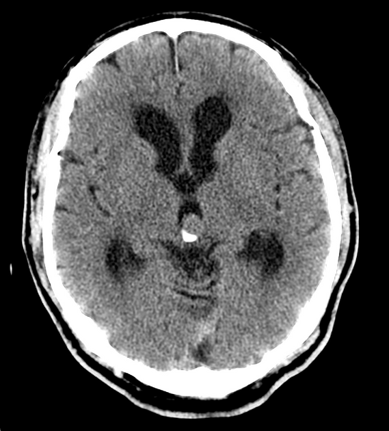
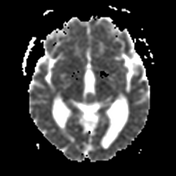
A 14 X 13 (axial) mm soft tissue density mass in the pineal region with eccentric peripheral calcification demonstrated. The ventricles are prominent, out of keeping with the degree of sulcal prominence, indicating mild to moderate obstructive hydrocephalus due to the lesion partially obstructing the upper margin of the cerebral aqueduct. Periventricular hypoattenuation is in keeping with chronic small vessel ischemia +/- transependymal CSF accumulation. No other significant intracranial abnormality.


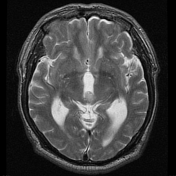
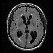
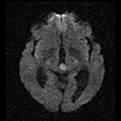
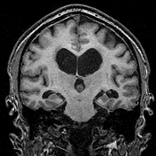
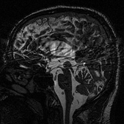
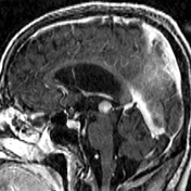
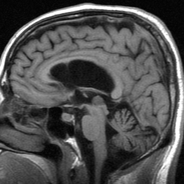
Circular mass centered in pineal region measures 12 x 11 x 9 mm and demonstrates homogeneous enhancement (on volumetric sequence for stereotaxis), peripheral calcification and diffusion restriction. It has mass effect on the adjacent structures with stenosis of the cerebral aqueduct (some flow still present on cine imaging) and associated non-communicating hydrocephalus affecting the lateral ventricles and 3rd ventricle. Periventricular FLAIR hyperintensity may represent some transependymal edema superimposed on chronic small vessel disease.
Foramina of Monroe are patent. No other suspicious enhancing lesion, including of the leptomeninges. No suprasellar/sellar mass in non-dedicated study.

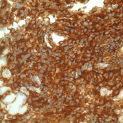
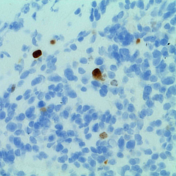
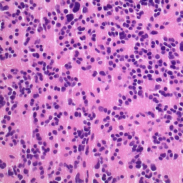
The sections show a moderately cellular tumor. It often forms rosettes and nests, surrounded by vascularized stroma. The tumor cells have generally round nuclei, granular chromatin, small nucleoli and moderate amounts of eosinophilic cytoplasm. No nuclear molding is seen. Occasional cells show degenerate nuclear changes with moderate nuclear enlargement and smudged chromatin pattern. No mitoses are seen. There is no endothelial cell hyperplasia or necrosis. The tumor cells are diffusely positive for synaptophysin and neurofilament. The Ki-67 index is 2-3%.
FINAL DIAGNOSIS: pineocytoma (WHO Grade I).
Case Discussion
Typical appearances of a small pineocytoma.


 Unable to process the form. Check for errors and try again.