
Presentation
Increasing headaches
Patient Data

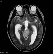
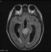
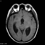
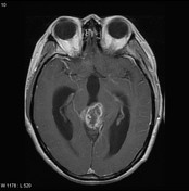


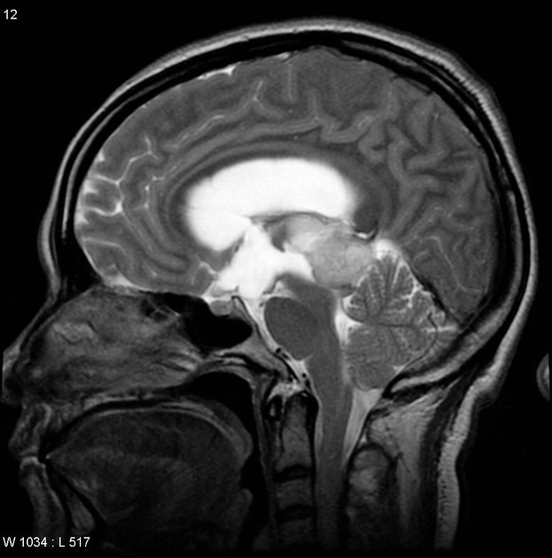
A large enhancing mass is present in the pineal region compressing the tectum and resulting in obstructive hydrocephalus.
The patient went on to have surgery and resection of the mass.
Histology
Sections show a tumor comprised of cells with small rounded nuclei with a moderate degree of pleomorphism. Some hyperchromatic nuclei are present, some of these with eosinophilic cytoplasmic inclusions. Cells show minimal cytoplasm but show elongated cell processes. In many areas the tumor shows anuclear zones. Mitoses are rare (there is only one seen in all of the sections). There is no endothelial proliferation and there is no necrosis. Immunohistochemistry shows that the tumor is strongly positive for synaptophysin and focally positive for neuron-specific enolase. Many of the tumor cells are positive for glial fibrillary acidic protein (GFAP).
Case Discussion
Path proven pineocytoma.


 Unable to process the form. Check for errors and try again.