
Presentation
Visual failure.
Patient Data
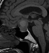
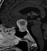
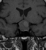
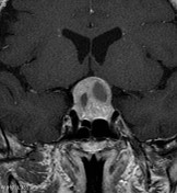

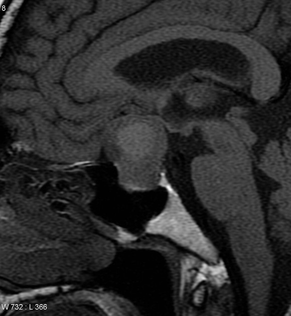
MRI demonstrates a large mostly solid enhancing mass enlarging the pituitary fossa and extending into the suprasellar cistern, elevating and compressing the optic chiasm. Central areas demonstrate cystic change with some intrinsic high T1 signal.
Case Discussion
Histology
Sections show multiple fragments of a pituitary adenoma, sclerotic stroma, and a few bony chips. The tumour cells are uniform and are laid in sheets and acini. The cytoplasm is amphophilic. There are no atypical features identified. The reticulin stain confirms the loss of acinar structure. No normal pituitary tissue is identified in the vicinity of the tumour. Immunohistochemistry for ACTH, prolactin, growth hormone, TSH, FSH and LH is negative.
Final diagnosis: Nonfunctioning (null cell) pituitary adenoma.


 Unable to process the form. Check for errors and try again.