
Presentation
Headache and increased prolactin levels.
Patient Data


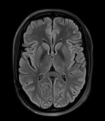
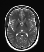
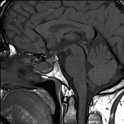
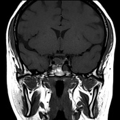
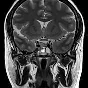
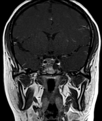
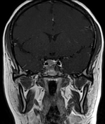
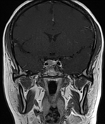
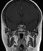
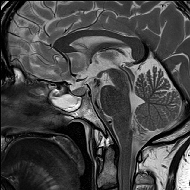
Focal lesion in the pituitary gland on right side measuring 8.6 x 9.4 x 8.5 mm, causing a bulge in superior contour of pituitary. It appears isointense on T1 and slightly hyperintense on T2 images. It is partially encasing the cavernous segment of right ICA with no obvious extension into the cavernous sinus. Mild expansion of sella turcica with no obvious extension into the sphenoid sinus. Mild deviation of the pituitary stalk towards left. No mass effect seen on optic chiasma.
On dynamic post contrast images, it enhances mildly against background of intensely enhancing normal pituitary.
Case Discussion
Findings are consistent with a pituitary microadenoma.
Lesions less than 10 mm are termed microadenoma. Those larger than 10 mm are termed macroadenomas.


 Unable to process the form. Check for errors and try again.