
Presentation
A pregnant woman comes for a routine ultrasound.
Patient Data

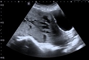

Images showed a total occlusive placenta with loss of the retroplacental clear space and reduced myometrial thickness. There are prominent vessels and lacunae through the placenta.
The color Doppler study shows increased vascularity and vascular bridges between the myometrium and the bladder wall.


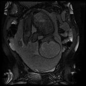
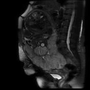
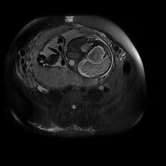
Magnetic resonance images showing the placenta covering the anterior wall, posterior wall, and the entire lower segment of the uterus, occluding the internal orifice of the cervical canal. T2 hypointense signal bands can be seen in the lower segment of the placenta adjacent to the body-cervix transition, with a lack of definition in the myometrium and signs of placental invasion in the entire thickness of the myometrium (placenta increta).
Additional findings: Maternal hydronephrosis.
Case Discussion
The placenta accreta spectrum (PAS) is characterized by abnormal adhesion and invasion of the trophoblastic tissue into the myometrium and uterine serosa and its invasion can be classified in placenta accreta (approximately 75% of cases), placenta increta as in the case, or placenta percreta.
The two most important risk factors are prior cesarian section and placenta previa.
Sonographic features of placenta accreta include loss of the normal retroplacental clear space, anomalies of the bladder- myometrium interface, prominent placental lacunae (highest positive predictive value), and increase vascularity at the interface uterus and bladder.
MR images are most useful in case of the sonographic findings are equivocal or when the placenta has a posterior location.


 Unable to process the form. Check for errors and try again.