
Plantar fasciitis, abductor digiti minimi atrophy and probable Baxter neuropathy
Presentation
History of heel pain.
Patient Data
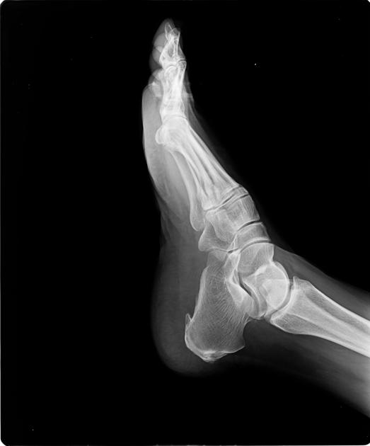
Plantar calcaneal spur is evident. Sclerosis and small spur is seen at posterior calcaneal tubercle.


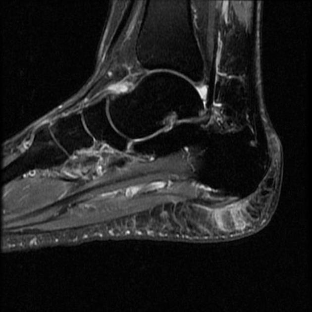
Saggital PD fat sat images exhibit mild thickening of the plantar fascia, predominantly lateral cord, with increased intrinsic signal, prominent plantar calcaneal spur, heel pad edema, and minimal focal subchondral edema at lateral calcaneal tuberosity, findings compatible with plantar fasciitis.
Coronal T1 images demonstrate fatty atrophy of abductor digiti minimi (ADMA).
Subchondral edema with degenerative cortical spurs seen at navicular - medial cuneiform joint.
The insertion of Achilles tendon shows minimal thickening with interlaminar fatty signal suggesting chronic tendinosis.
Minimal joint effusion is evident.
Case Discussion
MRI foot demonstrates calcaneal spur, plantar fascia thickening and edema, heel pad edema, abductor digiti minimi atrophy (ADMA), mild insertional tendinosis at Achilles - findings are consistent with chronic plantar fascitis with heel spur syndrome and ADMA likely consequence to inferior calcaneal nerve entrapment (Baxter's neuropathy)


 Unable to process the form. Check for errors and try again.