
Presentation
History of L5/S1 right-sided laminectomy and discectomy 14 months ago with persistent left S1 nerve root symptoms.
Patient Data


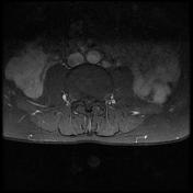
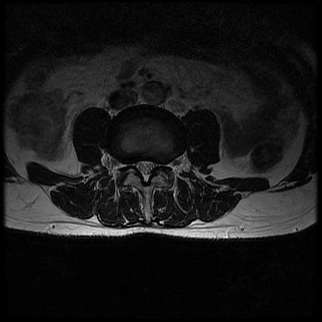
Axial T2-weighted MR image reveals an intermediate signal intensity lesion in the right epidural space including an ill-defined margin of the right S1 nerve root. Postcontrast axial T1-weighted image shows enhancement also extending to left epidural space, suggesting postoperative scar and focal enhancement of the thickened left epidural S1 nerve root .
Case Discussion
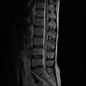
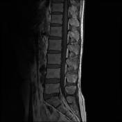
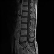
In patients with recurrent or residual sciatica after surgery for lumbar disk herniation, nerve root changes, especially root enhancement, on contrast-enhanced MR imaging six months postoperatively, showed the best association with clinical symptoms.


 Unable to process the form. Check for errors and try again.