
Presentation
posterior ankle pain
Patient Data
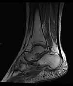

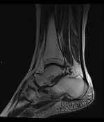
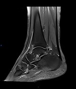
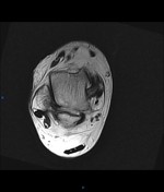
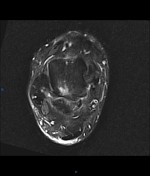
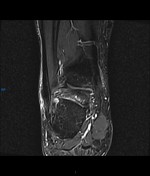
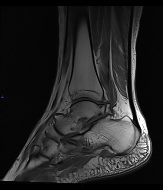
A large os trigonum with mild edema seen along the synchondrosis of an os trigonum and the talus with associated osteophytes, subarticular pseudocysts, and mild regional surrounding soft tissue edema. Osteophytes of the posterosuperior calcaneus opposed to the os trigonum are also noted.
Mild tibiotalar, talocalcaneal , and talonavicular osteoarthritic changes with marginal osteophytes
Minimal subcutaneous soft tissue edema posterior to the tendon Achilles
Minimal fluid distension of the flexor hallucis longus tendon sheath

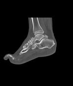
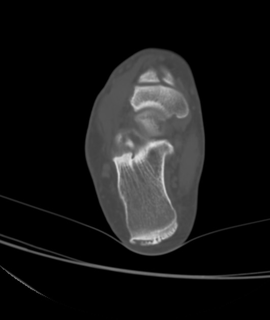
A large os trigonum with degenerative changes are seen along the synchondrosis of an os trigonum and the talus in the term of osteophytes and subarticular pseudocysts. Osteophytes of the posterosuperior calcaneus opposed to the os trigonum are also noted.


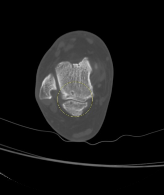
The yellow circles highlight the changes along the os trigonum/ talus synchondrosis
Case Discussion
Here is a case of Os trigonum syndrome, one of the predisposing factors of posterior ankle impingement with typical CT and MRI features along the os trigonum/ talus synchondrosis


 Unable to process the form. Check for errors and try again.