
Presentation
60-year-old lady presenting with significant parietal (dyspraxia, acalculia) and visuospatial and visuoperceptual problems (needs ruler to read, problems with stairs).
Patient Data
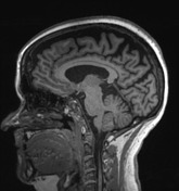

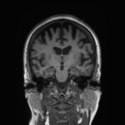
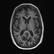
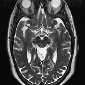
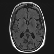

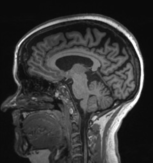
Normal grey-white matter differentiation. Volume loss in the parietal lobes, and possibly involving temporal lobes. The mesial temporal lobes including hippocampi are commensurate with the remainder of the brain. Only sparse chronic small vessel ischemic white matter changes are present, in a number acceptable for the age group. Ventricles are unremarkable. No extra-axial collections or mass are evident.
Conclusion: Presence of bilateral parietal lobe volume loss in the this age group and clinical setting favors posterior variant Alzheimer's disease as the most likely diagnosis.
Case Discussion
Volumetric segmentation shows markedly reduced parietal lobe volume, consistent with posterior cortical atrophy. The patient had gradual progression of her disease with a good response to acetylcholinesterase inhibitor, donepezil.


 Unable to process the form. Check for errors and try again.