
Presentation
55 year-old single woman with a PhD working up until last year. Admitted for investigation of approximately 2 year history of difficulty with decision making, spelling, writing, word-finding difficulties and dyspraxia.
Patient Data


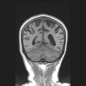
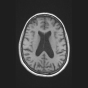

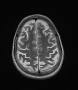
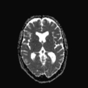
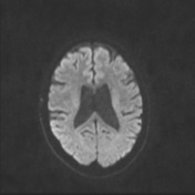
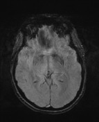
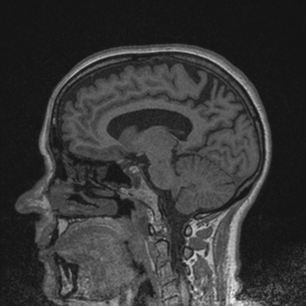
Bilateral marked parietal atrophy is demonstrates seen both on the interhemispheric and superolateral surface, involving precuneus and superior and inferior parietal lobules. The remainder of the brain is unremarkable in volume and appearance. The hippocampi are within normal limits. No susceptibility induced regions of signal loss to suggest microhemorrhages, no significant white matter T2 signal change and no abnormal restricted diffusion.
Conclusion:
Features are suggestive of posterior cortical atrophy (young onset posterior variant of Alzheimer's disease).
Case Discussion
Neuropsychology testing demonstrated profound impairment in non-verbal skills, profound slowing on non-verbal tasks ad marked impairment in aspects of verbal skills. Her memory was inefficient and slowed, with no rapid forgetting. Her performance on executive tasks was colored by her visuospatial impairment and processing speed. These findings are consistent with posterior cortical atrophy.
CSF demonstrated decreased levels of Abeta1-42, which is consistent with Alzheimer's Disease, as this protein is deposited and aggregated in amyloid plaques and hence decreased.


 Unable to process the form. Check for errors and try again.