
Presentation
4 days history of severe headaches, nausea and vertigo.
Patient Data
Age: 45 years
Gender: Male
From the case:
Posterior inferior cerebellar artery (PICA) infarct


Download
Info
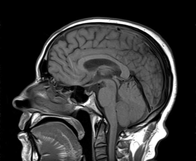
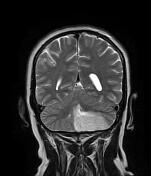
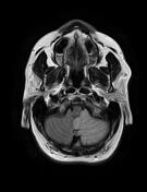
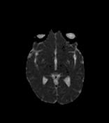
Large left cerebellar infarct involving the inferior posterior aspect of the cerebellum, the ipsilateral cerebellar tonsil and the lower part of the medulla oblongata (PICA territory). It appears as a large area of low signal on T1, high signal on FLAIR and T2 with restricted diffusion and no haemorrhagic component on the GE sequence. A mass effect is noted on the 4th ventricle.
Case Discussion
The clinical presentation and the MRI features are most consistent with a posterior inferior cerebellar artery (PICA) infarct.


 Unable to process the form. Check for errors and try again.