
Presentation
Abdominal pain, jaundice and fatigue
Patient Data

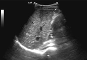
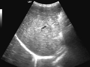
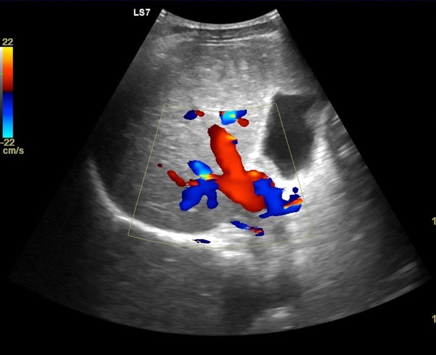
Ultrasound study of the liver shows coarse inhomogeneous hepatic parenchyma with irregular contour. Duplex study reveals normal vascularity of the portal vein.
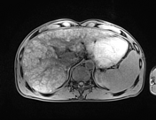

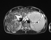
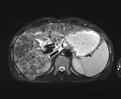
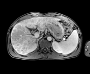
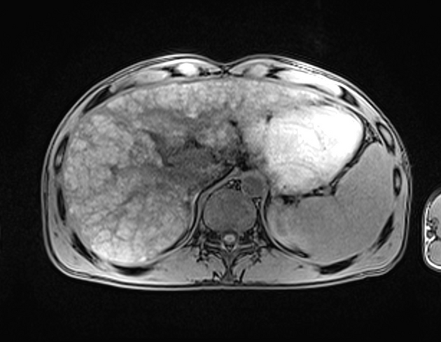
Average sized liver presents markedly heterogeneous parenchyma with nodularity and irregular contour. It elicits heterogeneous high signal on T1WI and T2WI with parenchymal "lace-like pattern". Periportal hyperintensity on T2WI, denoting periportal edema. Distended gallbladder with pericholecystic fluid.
Case Discussion
Primary biliary cholangitis (PBC) is a chronic progressive cholestatic liver disease that is characterized by the destruction of small intrahepatic bile ducts, portal inflammation, and progressive scarring. The cause of PBC is unknown, but it is probably due to an inherited abnormality of immunoregulation. It constitutes the third most common indication for liver transplantation in adults.
The name of this disease was changed from "primary biliary cirrhosis" to "primary biliary cholangitis" in 2014 3. Patients present with jaundice and high serum bilirubin. Serum anti-mitochondrial antibody (AMA) tests are highly sensitive and specific for PBC (85-100%). In this case, serum total bilirubin was very high (15.6 mg/dL) and anti-nuclear antibody was positive.


 Unable to process the form. Check for errors and try again.