
Presentation
Confusion and right sided weakness.
Patient Data

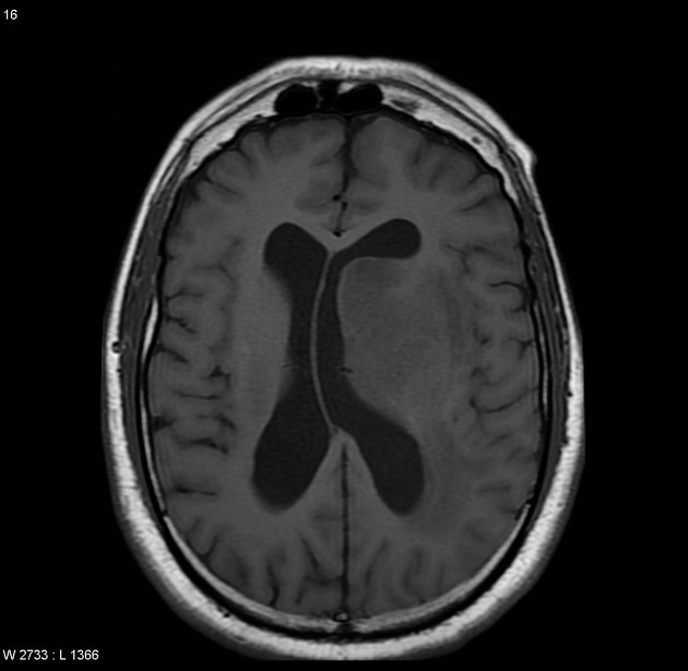
Non-contrast CT demonstrates a number of hyper-attenuating masses adjacent to the left lateral ventricle, with no calcification or hemorrhage.


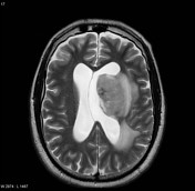
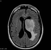
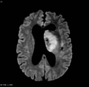
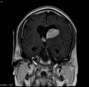
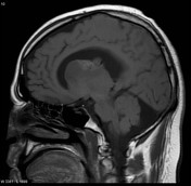
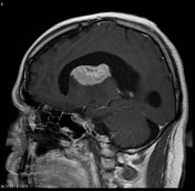
MRI confirms the masses, which are of high T2 and low to intermediate T1 signal. A moderate amount of vasogenic edema extends away from these masses into the white matter. The lesions demonstrate prominent restricted diffusion on DWI and intense homogeneous contrast enhancement.
This patient went on to have a stereotactically guided biopsy (as the diagnosis of lymphoma was strongly suspected on imaging), and this confirmed the diagnosis: high grade B-cell lymphoma.
Case Discussion
This case demonstrates features characteristic of CNS lymphoma (primary in this case), and highlights the importance of understanding the general histological makeup of lesions if imaging is to be adequately interpreted. The densely cellular and 'small round blue cell' appearance manifests as hypderdensity on CT and restricted diffusion on MRI, with out the heterogeneity or non-enhancing components of glioblastomas.
Although this is not an intra-ventricular lesion, the striking periventricular distribution and exophytic thalamic component means that it is useful to think of this lesion in the context of intraventricular pathology, which can often (e.g. ependymoma) have a parenchymal component.


 Unable to process the form. Check for errors and try again.