
Presentation
Previously healthy patient presents with somnolence, lethargy, and emotional lability over the past week.
Patient Data


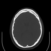

Axial unenhanced CT of the head demonstrates a lobular hyperdense mass in the floor of the third ventricle. There is subtle associated hypodensity in the adjacent optic tracts, suggestive of edema.

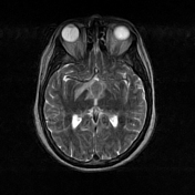
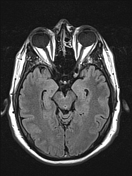


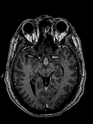


MRI of the brain displays an ovoid, fairly homogeneous mass centered in the third ventricle with extension into the optic recess. The mass is hypointense on T1-weighted imaging and mildly hypointense on T2-weighted imaging. Diffusion weighted images are mildly increased though diffusion coefficient is similar to brain parenchyma. The T2 signal and mild diffusion restriction in combination with hyperdensity on CT suggest increased cellularity. Following contrast administration, the mass displays homogeneous enhancement.
There is also increased signal on T2/FLAIR weighted images involving the optic tracts, chiasm, ventral midbrain around the mammillothalamic tracts, as well as around the anterior commissure, likely representing edema.
Case Discussion
The mass was surgically removed and pathologically proven to be diffuse large B cell lymphoma. This case represents characteristic imaging features of primary CNS lymphoma in an atypical location. This patient had no evidence of involvement of lymphoma outside the CNS including a negative bone marrow biopsy.


 Unable to process the form. Check for errors and try again.