
Presentation
Seizure.
Patient Data


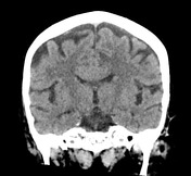

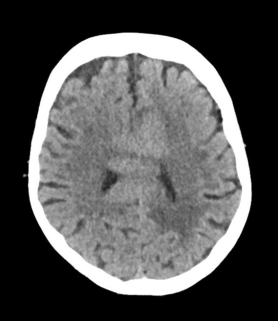
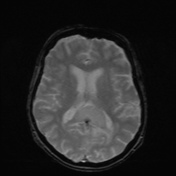
There are homogeneously hyperdense masses involving the body and splenium of the corpus callosum, crossing through both hemispheres, but mostly on the left. They appear somewhat confluent and are associated with mild white matter vasogenic edema on the left. The brain parenchyma is otherwise unremarkable. The ventricles and basal cistern are unremarkable. There is no cerebral herniation.
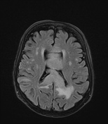
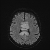

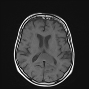

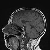
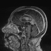
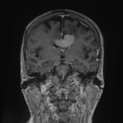
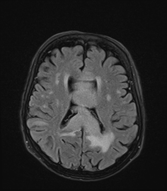
The callosal tumor has homogeneous and vivid contrast enhancement and also shows marked diffusion restriction.
Case Discussion
Typical epidemiology and imaging features of PCNSL.
Macroscopic: A. B. C. Labeled "Corpus callosal lesion"
Microscopic: A. B. C. The tissue is cellular, showing infiltration by large atypical lymphoid cells with frequent apoptosis. Cell nuclei show weak pleomorphism and small nucleoli and atypical mitoses. Cells are immunoreactive with CD20, BcL2 (100% cells), BcL6 (60% cells), Ki67 in 100%. Cells are negative for CD3, CD 30, and EBV.
Diagnostic opinion: A. B. C. Corpus callosal lesion – high-grade B cell lymphoma.


 Unable to process the form. Check for errors and try again.