
Presentation
Painful right shoulder.
Patient Data
Age: 16 years
From the case:
Primary synovial osteochondromatosis

Download
Info

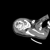
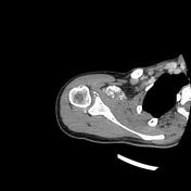
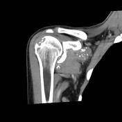
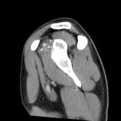
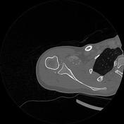
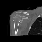
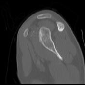
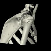
The CT scan demonstrates multiple small calcific loose bodies of uniform size within the glenohumeral joint, axillary recess, subacromial/subdeltoid bursa as well as the subscapular bursa and the biceps tendon sheath. Irregular destruction of the posterolateral aspect of the humeral epiphysis containing chondroid calcifications. Irregular cortical erosions of the glenoid cavity are also noted. No evidence of osteoarthritic changes.
Case Discussion
The patient age, clinical presentation and CT features are most consistent with a primary synovial osteochondromatosis.


 Unable to process the form. Check for errors and try again.