
Presentation
Metrorrhagia with a mass visible through the vaginal orifice on the gynecological examination.
Patient Data
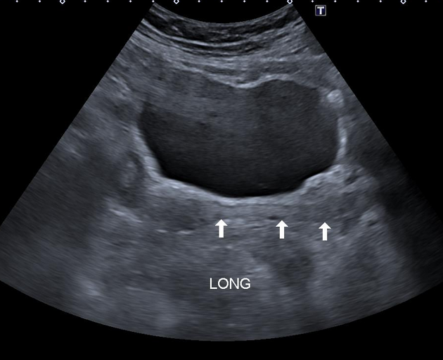
Normal size and echotexture of the uterus. Echogenic mass fills the uterine cavity and extends through the endocervical canal filling the entire vagina (white arrows).


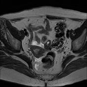
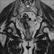
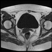
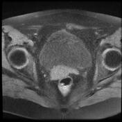
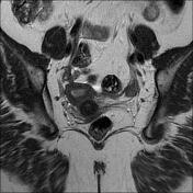
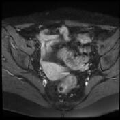
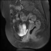
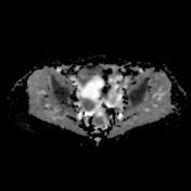
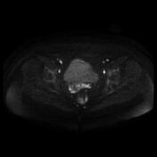
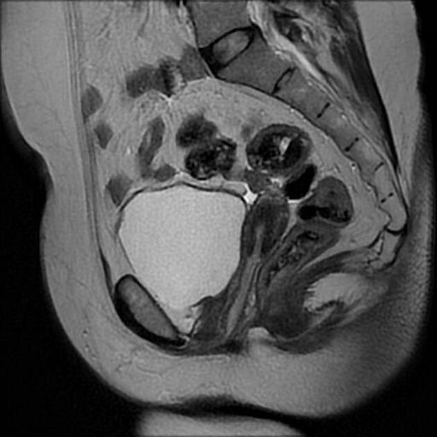
Well-defined pedunculated intracavitary soft tissue mass (length = 10 cm), arising from the fundal wall, prolapsing into the entire vaginal canal through a moderately enlarged endocervical canal. It displays an isosignal on T1, intermediate to high signal on T2 with surrounding hyperintense endometrium and no restricted diffusion. The postcontrast sequences show a heterogeneous enhancement. No infiltration of the myometrium. No pelvic lymphadenopathy is seen. Minimal effusion in Douglas pouch and in inter vesicouterine space.
Case Discussion
Ultrasound and MRI features most consistent with an endometrial polyp prolapsed into the entire vaginal canal.


 Unable to process the form. Check for errors and try again.