
Presentation
Orthopedic clinic follow up.
Patient Data
Age: 30 years
From the case:
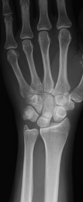
Proximal row carpectomy

Download
Info
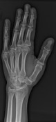
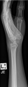
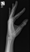
Removal of the scaphoid, lunate and triquetral bones in keeping with proximal carpectomy. The proximal capitate is articulating with the lunate fossa of the distal radius as expected. Alignment is stable when compared to the prior study.

Download
Info
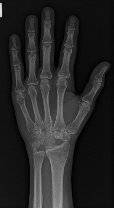
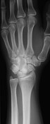
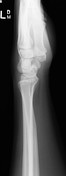
On the frontal view there is widening of the scapholunate interval with decreased radioscaphoid joint space. The lunate has a triangular configuration. The lateral view shows dorsal subluxation of the scaphoid, dorsal tilt of the lunate and increased scapholunate angle consist with DISI.
Case Discussion
The patient had severe pain and instability after a untreated scapholunate ligament tear progressed to SLAC and was treated successfully with PRC.


 Unable to process the form. Check for errors and try again.