
Presentation
Persistent pain under the foot for a few months.
Patient Data
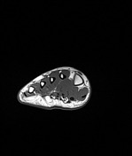

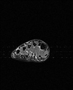
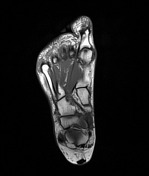
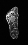
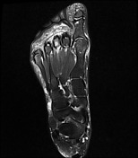
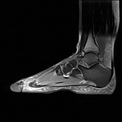
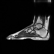

Focal discontinuity of the plantar plate of the 3rd metatarsophalangeal joint associated with abnormal soft tissue signal around the plantar plate extending into the adjacent intermetatarsal space is seen, which unlike a true neuroma shows ill-defined contours and has an asymmetric position with respect to a vertical line drawn through intermetatarsal space.
As well, degenerative changes at the 1st metatarsophalangeal joint associated with cystic at the subjacent portion of the distal portion of the 1st metatarsal bone, edema and irregularity at medial sesamoid of great toe. Mild filling of the 2nd intermetatarsal bursa, suggestive of small Morton’s neuroma is also seen. Foci of plantar soft tissue fibrosis/edema at the level of the 1st and 5th metatarsal heads, suggestive of callus formation/mild adventitial bursitis are also noted.

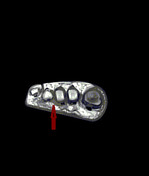
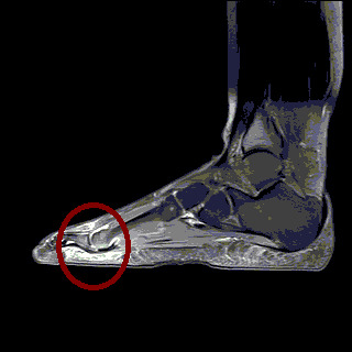
The place of plantar plate tear and associated fibrosis (pseudoneuroma sign)
Case Discussion
Two strongly associated indirect signs of plantar plate tear are the psedoneuroma sign and plantar plate-proximal phalanx distance (>0.275 cm).
In comparison with Morton neuroma, pericapsular fibrosis (pseudoneuroma sign) presents as an ill-defined and eccentric soft tissue thickening with the sequential progression of enthesitis, flexor tendon subluxation and splaying of the toes is helpful for optimizing accurate diagnosis of plantar plate tear and associated pericapsular fibrosis.


 Unable to process the form. Check for errors and try again.