
Presentation
Presenting with chronic pelvic/groin pain. No history of trauma. Clinical concern for pudendal neuralgia.
Patient Data


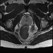
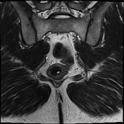
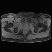
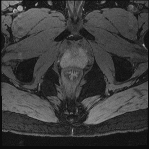
MRI shows the course of the pudendal nerve at the greater sciatic foramen, at the ischial spine, at the lesser sciatic foramen, and in Alcock's canal. The terminal branches include the inferior rectal nerve, perineal nerve, and dorsal nerve of the penis (in males). The major branch of the perineal nerve is the posterior scrotal nerve (in males). The sacrospinous ligament and sacrotuberous ligament appear normal. The sacrospinous ligament separates the greater sciatic foramen from the lesser sciatic foramen. The piriformis muscle and gluteus maximus muscle appear normal.
The unique anatomy of the pudendal nerve is noted as it exits the pelvis via the infrapiriform greater sciatic foramen but then re-enters the pelvis via the lesser sciatic foramen. The sciatic nerve exits at the greater sciatic foramen and continues laterally.
The prostate and urinary bladder appear normal.
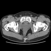
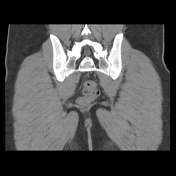
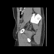
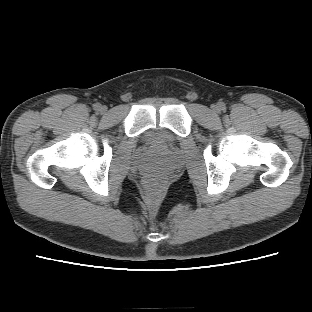
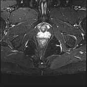
For comparison, CT from the same patient shows the course of the pudendal nerve at the greater sciatic foramen, at the ischial spine, at the lesser sciatic foramen, and in Alcock's canal.
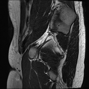
There is oral contrast in the bowel. Chronic bilateral pars defects are present at L5.
Case Discussion
Pudendal neuralgia secondary to pudendal nerve entrapment is a debilitating condition that is most commonly diagnosed clinically 1. With the advancement of modern imaging technology, it is possible to visualize the pudendal nerve on MRI 2.
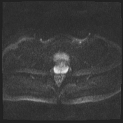
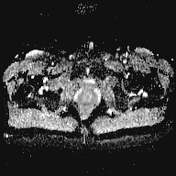
Thickening and hyperintensity of the affected pudendal nerve can be observed in pudendal neuralgia, especially when compared to the contralateral pudendal nerve. The Double Echo Steady State (DESS) sequence provides high resolution images for morphologic nerve assessment. It is important not to mistake adjacent vessels for abnormal nerve signal.
MRI allows for radiological correlation to patient presentation/symptoms. Whereas pudendal neuralgia has traditionally been diagnosed clinically with the Nantes criteria 3, MRI may now be used to aid in diagnosis.


 Unable to process the form. Check for errors and try again.