
Presentation
Pelvic pain with vaginal discharge.
Patient Data

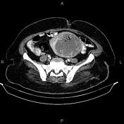
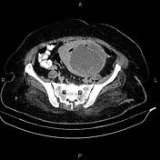
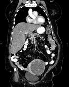
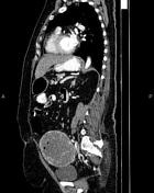

The uterus is enlarged. A 115×100×95 mm cysts like lesion with internal air bubble is seen at uterus body. Some small air bubbles are also evident at endometrial cavity.
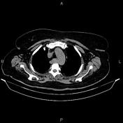
The right subclavian artery is arising from the arch of aorta directly posterior to the esophagus inferring aberrant right subclavian artery.
The hepatic attenuation value is less than of the spleen, suggesting fatty liver disease.
An 18 mm thin walled non enhanced cyst is noted at pancreatic tail.
A few sub centimeter simple cortical cysts are seen at kidneys. A 15 mm stone (476HU) is present at right renal pelvis. A 9 mm stone (420HU) is also evident at right renal middle calyx. Additionally, a few stones less than 4 mm are observed at left renal.
Fat containing para umbilical hernia is present.
Degenerative changes as osteophytosis are seen at the lumbar spine.
Grade I spondylolisthesis of L4 on L5 is present.
Case Discussion
Large uterus cyst like lesion most consistent with necrotic uterine leiomyoma with superimposed infection also known as pyomyoma. The patient underwent surgical resection and pathologically confirmed.


 Unable to process the form. Check for errors and try again.