
Presentation
Pituitary cystic lesion incidentally found in a previous brain MRI.
Patient Data
Age: 45 years
Gender: Male
From the case:
Rathke cleft cyst


Download
Info
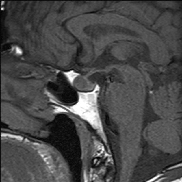
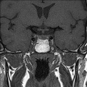
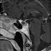
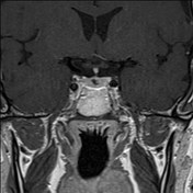
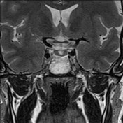
Cystic lesion located in the midline of the adenohypophysis that appears hyperintense in T2 sequence and isointense in T1 enhanced sequences.
It measures 10 x 8 x 8 mm, has a thin wall that enhances with contrast and a 5mm bilobulated T2 hypointense non-enhancing intralesional nodule, which is pathognomonic for Rathke cleft cyst.
It does not affect to the optic chiasm.
Case Discussion
A cystic hypophyseal lesion with a characteristic small non-enhancing intracystic nodule. Findings are consistent with Rathke cleft cyst.


 Unable to process the form. Check for errors and try again.