
Presentation
Non-traumatic pain in right proximal medial leg. Recent radiograph was normal. Patient was referred for local part ultrasound.
Patient Data

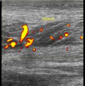
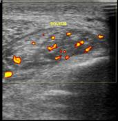
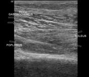
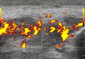
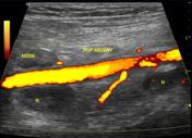
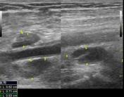
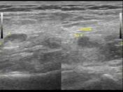
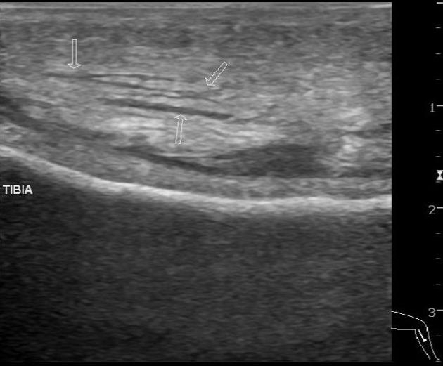
Edematous area of the proximal belly of soleus muscle, distal belly of popliteus muscle is noted with adjacent soft tissue hypoechogenicity and hypervascularity. No collection is noted.
No obvious erosion of adjacent tibial cortex is noted.
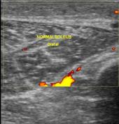
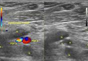
Reactive (non-necrotic and ovoid) popliteal nodes are seen in the popliteal fossa. Five in number.
Case Discussion
Local infectious/inflammation involving soleus, popliteus muscles and adjacent soft tissue was suggested on ultrasound without collection (no abscess formation) without tibial cortical erosion.
MRI was done to look for marrow signal abnormality in tibia; which was normal. A rim of subperiosteal fluid was noted.
Popliteal lymphadenopathy can be due to infection (as in this case and case nº 13658), melanoma (case nº 25965) or due to various other causes.


 Unable to process the form. Check for errors and try again.